The Fascinating World of MRI: How It Works and Why It Matters
Decoding the Mystery: What is MRI Technology?
Magnetic Resonance Imaging (MRI) is a non-invasive diagnostic technique that provides detailed images of internal body structures, particularly useful for examining bones and joints. Unlike X-rays or CT scans, which utilize ionizing radiation, MRI employs powerful magnets and radio waves to generate images. The primary component of MRI technology is its magnet, which can be several times stronger than the Earth’s magnetic field. When a patient enters the MRI machine, the magnet aligns the protons in their body—mostly found in water molecules—creating a magnetic field. When radiofrequency pulses are applied, these protons are knocked out of alignment, and as they return to their original position, they emit signals that a computer captures and converts into images.
MRI technology has evolved significantly since its inception, with advancements in sequence acquisition, gradient strength, and software leading to increasingly high-resolution images. It is particularly beneficial for visualizing soft tissues, including muscles, ligaments, and cartilage, making it an essential tool for diagnosing and evaluating musculoskeletal conditions. Understanding MRI technology not only demystifies the process but also highlights its significance in providing healthcare professionals with essential insights for effective treatment planning.
Exploring the Science: How MRI Captures Bone and Joint Images
The science behind bone and joint MRI is rooted in the difference in tissue properties within the body. When the protons realign after being disturbed by radiofrequency waves, the time it takes varies between different types of tissues, which is critical for creating the contrast seen in MRI images. Typically, fluid-rich tissues, such as cartilage and synovial fluid, appear brighter compared to denser, less hydrated tissues like bone, which often shows as a darker area on scans. This contrast is instrumental in diagnosing a plethora of conditions, as it allows radiologists to distinguish between healthy tissues and those affected by disease or injury.
In addition, advanced MRI techniques such as diffusion-weighted imaging or magnetization transfer imaging can provide even more actionable data. For instance, diffusion-weighted imaging is excellent for assessing edema and the biochemical microenvironment surrounding an injury, while magnetization transfer imaging offers insights into cartilage integrity. Such detailed imaging capabilities are invaluable, as they can reveal the subtle changes in bone and joint structures before they become clinically apparent, allowing for earlier intervention and better patient outcomes.
The Role of Contrast Agents: Enhancing Visibility for Better Diagnosis
In many cases, a standard MRI may not provide sufficient detail for a comprehensive diagnosis. This is where contrast agents come into play. These substances, usually gadolinium-based, enhance the visibility of certain tissues by altering their magnetic properties. When injected into a patient, contrast agents distribute through the bloodstream and can highlight areas of inflammation, tumors, or infections that might be indistinguishable in non-contrast images. The use of contrast in MRIs is particularly beneficial when evaluating complex joint structures, where conditions like synovitis or osteomyelitis need to be examined thoroughly.
However, the use of contrast agents is not without risks. Some patients may have allergic reactions or experience nephrotoxic effects, particularly those with pre-existing kidney issues. As such, clinical discretion should be exercised, and thorough historical evaluation of kidney function is required before administering contrast during an MRI exam. Despite these risks, the benefits gained from enhanced imaging often outweigh the concerns, facilitating more accurate diagnoses and leading to improved health management strategies.
Common Bone and Joint Conditions: What MRI Reveals
From Arthritis to Fractures: Understanding Different Conditions
MRI plays a pivotal role in diagnosing various bone and joint conditions, from degenerative diseases like arthritis to traumatic injuries such as fractures. Osteoarthritis (OA), the most common form of arthritis, is characterized by the wear-and-tear of cartilage, leading to joint pain and stiffness. MRI can detect early changes in cartilage and the surrounding area, helping to differentiate OA from other forms of arthritis, such as rheumatoid arthritis (RA), which involves inflammation of the synovium. Knowledge of these varying conditions allows for tailored treatment plans that can significantly alleviate symptoms and improve quality of life.
Moreover, fractures can manifest differently in MRI depending on the timing of the scan. Acute fractures may present as bright areas on an MRI due to associated edema or soft tissue damage. Conversely, stress fractures, caused by repetitive strain, may not always be visible on X-rays but can be accurately identified on MRI, allowing for appropriate rest and recovery recommendations. Timely diagnosis of fractures through MRI can prevent further damage and impeding recovery.
Soft Tissue vs. Hard Tissue: What MRI Can Show You
The distinction between soft and hard tissues in MRI imaging exemplifies the technology’s versatility. MRI is incredibly proficient at visualizing soft tissues—muscles, tendons, ligaments, cartilage, and nerves—due to their water content, which creates a strong contrast against surrounding muscles and fat. In contrast, MRI is less effective for seeing dense hard tissues like bone, as they generally appear dark and can obscure important details. However, the ability to assess the interface of soft and hard tissue, as well as the surrounding soft tissue structures, is invaluable in providing a comprehensive view of bone and joint health.
Pathologies such as tears in the rotator cuff or ligaments, bone marrow edema, and conditions like tendinopathy can be accurately visualized with MRI, guiding effective treatment approaches. For instance, in the context of a meniscal tear in the knee, MRI findings can reveal the tear’s size and location, providing critical information for potential surgical intervention planning. The thorough understanding of both soft and hard tissues is essential for consolidation in the diagnosis of sport-related injuries and chronic conditions, ensuring optimal care for patients.
Red Flags in MRI Findings: When to Seek Further Evaluation
While MRI is an incredibly powerful diagnostic tool, interpreting its findings requires a keen understanding, particularly regarding red flags that indicate the need for immediate further evaluation. Abnormal growths, bone lesions, or extensive edema can signify underlying malignancies, infections, or severe degenerative diseases and warrant rapid follow-up care. MRI findings of the spine, for example, can reveal congenital anomalies or degenerative disc disease that might lead to significant morbidity if left untreated.
Alongside direct findings, indirect indicators such as the presence of inflammation or suspicious changes in adjacent soft tissues can provide critical context for determining the urgency of clinical intervention. Healthcare professionals must educate patients about these potential red flags in their MRI report, as timely referrals and targeted therapies can vastly improve outcomes, especially in the face of serious conditions like tumors or infections. In summary, awareness of such red flags and understanding the implications of MRI findings is crucial for ensuring that health concerns are addressed proactively.
Preparing for Your MRI: Tips and Considerations
What to Expect: The MRI Experience Demystified
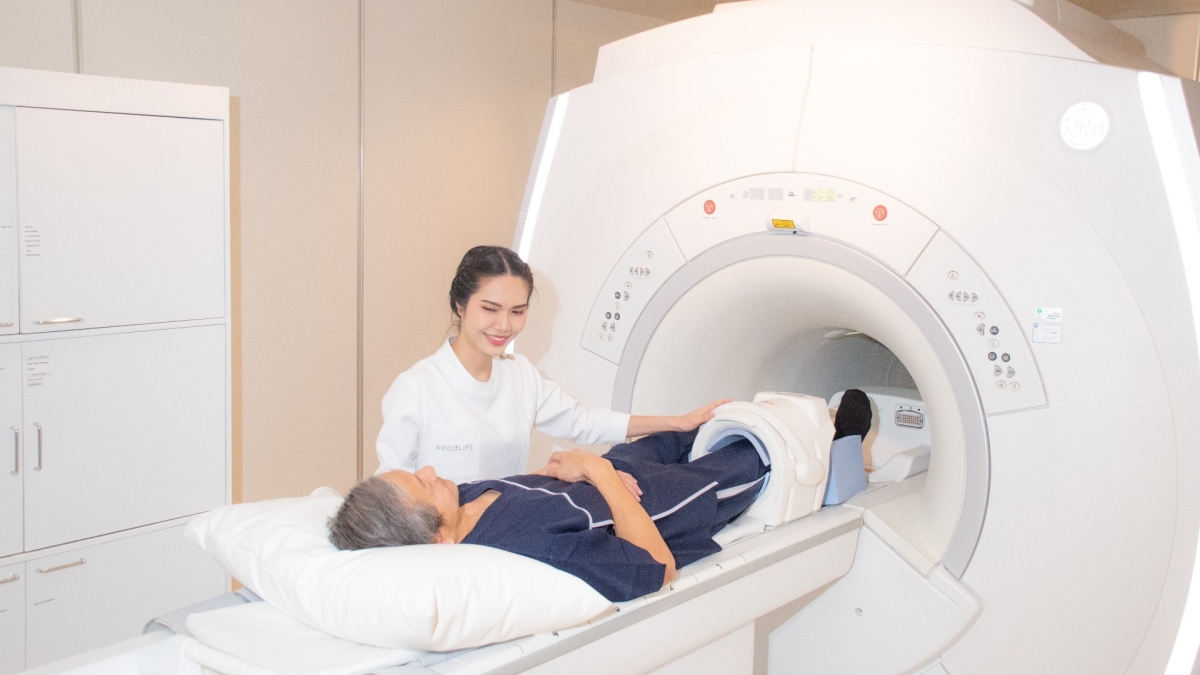
Facing your first MRI can feel intimidating, but understanding the procedure can alleviate anxiety and set realistic expectations. Upon arrival at the imaging center, you will be asked to complete necessary paperwork, after which a radiologic technologist will guide you through the process. Preparation often involves a brief medical history review to check for any contraindications, especially if you have meta devices or an allergy to contrast media.
Once you are ready, you will be positioned on a padded table that slides into the MRI machine, which resembles a large tube. It’s important to remain still during the scanning process, which can last anywhere from 15 minutes to over an hour, depending on the specific scans ordered. As the machine generates loud tapping or thumping noises, earplugs or headphones may provide comfort and minimize any anxiety related to the sounds. Some facilities may also offer visual distractions, such as screens displaying calming imagery. Overall, approaching the MRI with understanding and knowledge can convert what seems like a daunting experience into a straightforward and manageable one.
Clothing and Accessories: Dress Smart for Your Scan
When preparing for your MRI, careful attention to clothing and personal items can significantly enhance the efficiency of the process. Comfortable, loose-fitting clothing is often recommended, ensuring that no metal is present, as metal can interfere with MRI imaging. Patients may be asked to change into a hospital gown, which allows for easy access to the area being imaged, minimizing potential movement disruptions. Jewelry, watches, or any metallic hairpins must be removed before the procedure; even small items can create artifacts on the images that could compromise the effectiveness of the scan.
Additionally, consider personal comfort—if you require mobility aids, such as canes or walkers, verify that they are MRI-safe or, if possible, make arrangements to have someone assist you through the process. Overall, embracing a thoughtful approach to clothing and accessories will streamline your MRI experience while ensuring maximal comfort and limit unnecessary delays in the scanning process.
Addressing Concerns: Claustrophobia and Anxiety Management
For many individuals, the idea of being enclosed in an MRI machine can provoke feelings of claustrophobia or anxiety. It’s crucial to communicate any such apprehensions with your healthcare provider prior to the procedure so that measures can be taken to ease your concerns. Facilities frequently employ open MRI machines—designed to offer a more spacious experience—that can accommodate those uneasy in traditional closed units. If open MRI is not an option, virtual reality headsets or guided imagery techniques may also be employed to offer distractions during the scan.
Additionally, some patients may benefit from medication that alleviates anxiety before an MRI. It is essential to discuss this option with your doctor, who can prescribe appropriatesedatives, allowing you to remain calm and relaxed during the scan. Many facilities also promote the use of calming techniques such as deep-breathing exercises, which can significantly reduce feelings of anxiety and discomfort before the procedure starts. Ultimately, addressing these concerns beforehand can turn the MRI experience into a more tolerable one, resulting in more accurate imaging outcomes.
Interpreting MRI Results: Insights from Radiologists
The Art of Reading MRI Images: What Radiologists Look For
The interpretation of MRI images requires a unique blend of clinical knowledge and expert training. Radiologists look for specific patterns and inconsistencies within the images, assessing not only structural changes but also the relationship between various tissues. They analyze the characteristics of various tissues, noting signal intensities, which vary depending on hydration levels, tissue type, and pathology. Furthermore, radiologists often utilize advanced imaging techniques, like multi-sequence protocols that provide an array of views to accurately diagnose complex conditions.
Radiologists engage in a methodical process, examining images from different angles and perspectives while cross-referencing historical patient data and presenting symptoms. A sophisticated understanding of anatomy, pathology, and the implications of observed changes allows radiologists to make informed conclusions, leading to precise diagnoses and tailored treatment recommendations. Continuous education and training are crucial for radiologists, as advancements in MRI technology and techniques evolve rapidly, necessitating an up-to-date understanding to provide the highest level of care.
Understanding Reports: Breaking Down MRI Terminology
Receiving your MRI report can be daunting, as medical jargon often obscures the significance of findings. Familiarizing yourself with common terms can facilitate a more productive discussion with your healthcare provider. For instance, terms like “edema” indicate the presence of excess fluid, suggesting inflammation or injury, while “tear” refers to damage in soft tissue structures like tendons or ligaments. Knowing the distinction between “hypointense” and “hyperintense” signals can also be key; the former describes areas appearing darker on images (often denser tissues) while the latter indicates lighter areas (often reflective of fluid or inflammation).
It is also essential to understand that not every finding in an MRI is a cause for concern. Conditions like degenerative changes may be noted, but these can be age-related and not necessarily symptomatic. Engaging your healthcare provider in an open dialogue regarding your MRI results is critical, ensuring you gain a thorough understanding of your body’s condition and potential treatment pathways.
Follow-Up Care: Next Steps After Your MRI Exam
After your MRI exam, the next steps hinge on the interpretive findings and the specific condition that has been evaluated. Follow-up care may involve further diagnostic tests, referrals to specialists, or initiating treatment protocols based on the results. For instance, in cases where a significant injury or disease is detected, consulting with orthopedists, rheumatologists, or physical therapists may be appropriate. Individuals may also need to undergo follow-up imaging to monitor the progression of a condition.
Patient education plays a vital role in the follow-up phase; understanding your MRI findings equips you to make informed decisions regarding treatment options. This may include engaging in rehabilitation sessions, lifestyle modifications, or exploring surgical interventions based on the findings. Regardless of the pathway, receiving this information can empower you to take control of your healthcare journey, facilitating the best outcomes for your condition. In conclusion, MRI stands as an indispensable tool in understanding bone and joint health, and comprehensive interpretation of both images and reports can significantly enhance patient care.
Laila Azzahra is a professional writer and blogger that loves to write about technology, business, entertainment, science, and health.